Why does buttock pain flip from side to side in AS?
Introduction
A major feature of Ankylosing Spondylitis (AS) is deep seated buttock pain that flips from side to side. Frustratingly, when the doctors examine a patient with such pain, there may be nothing to find. This contributes to a delayed or incorrect diagnosis. This page explains the nature of such pain in terms of enthesis, bone and the mechanical stressing theory of AS.
Mechanism of alternating buttock pain
The sacroiliac joint is lined by fibrocartilage. This tissue is also present at enthesis attachment points to bone.
Fibrocartilage is subject complex patterns of mechanical stressing and this is transmitted to the underlying bone. This bone stressing and possible associated microdamage leads to bone inflammation (osteitis) that results in pain that is typical of AS.
Once an area of microdamage is repaired the pain may disappear. An abnormal response to bone stressing or regions of microdamge may randomly occur on the opposite sacroiliac joint. As a result the pain may come back on the opposite side. Ongoing cycles of inflammation flipping from side to side may lead to a pattern of alternating buttock pain.
Why doctors may not recognise sacroiliitis in subjects with buttock pain
First the joint moves very little so the best test of arthritis, which is limitation of movement, does not really apply.
Secondly the joint is deeply located to large muscles around the pelvis. The part of the joint that is inflamed is usually the front of the joint which is completely inaccessible to the examiners hands.
Third the inflammation is within the bone or what is termed trabecular bone. The tough outer shell of the bone, termed the bone cortex is not usually involved and when doctors press on this no tenderness is elicited.
The blood tests that usually confirm inflammation may be negative because the region of inflammation is relatively small being confined within a small region of bone.
The inflammation may take time to lead to bone damage so X-rays may be normal in early disease or even indefinately.
Illustrative case
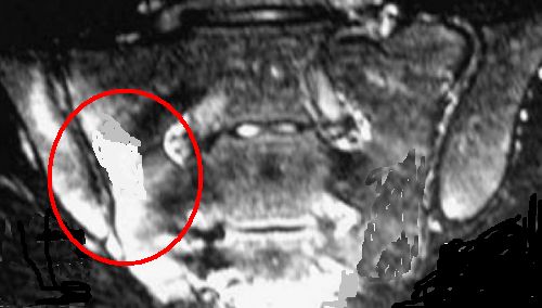
A young male patient who was HLA-B27 positive came to medical attention with inflammatory back pain that was felt in the right buttock region. The initial MRI scan is shown below.
|
| This MRI scan shows severe "bone oedema" on the fat suppressed MRI scan of the right sacroiliac joint. The bone oedema corresponds to bone inflammation (osteitis) and is located inside the red circle |
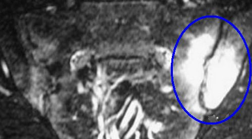
Approximately 6 months later the patient felt pain in the left buttock. The clinical examination elicited no tenderness. The second MRI scan is shown below and shows how the bone inflammation has flipped to the other side of the joint.
|
| The second MRI scan was performed 6 months later when the patient now had severe pain in the left buttock but none in the right. The inflammation in the bone is located inside the red circle. This is located deep to the buttock muscles. |
Implications
Alternative buttock pain is suggestive of early sacroiliitis. This pain is sometimes incorrectly given the wrong diagnosis of sciatica. The buttock pain related to sacroiliitis may go as far as the knee but generally no further.
Finally, inflammation may start elsewhere in the SIJ so not everyone gets alternating buttock pain.
References

Resources
Arthritis Research UK Spondyloarthritis