Ultrasound of Damage in Normal Insertions
Introduction
Tissue studies of the enthesis from elderly deceased subjects have shown that microscopic bone cracks, microscopic bone erosions, microscopic fibrocartilage damage, abnormal blood vessel formation and microscopic inflammation are very common. It is not possible to obtain such tissue from young healthy people who are physically very active. This page shows examples of damage at the normal enthesis as detected by ultrasound scanning.
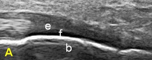
|
| this is an ultrasound image of a completely normal Achilles enthesis (e). The bone is marked (b). The black region on the bone surface is normal stress resisting fibrocartilage (f). |
Examples of damage at Normal Insertions
The fibrocartilage is a stress shield at the normal insertion. This gets damaged in disease but also shows subtle damage even in normal subjects [1]
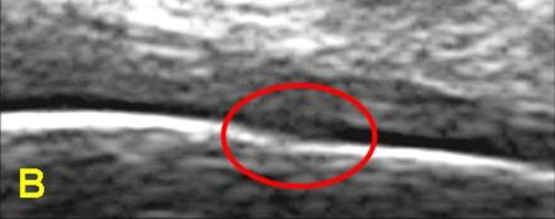
|
| this is an ultrasound image from a healthy young man without any heel pain showing microdamage. The normal black colour at the site of fibrocartilage is lost and likely represents microdamage at that site. |
Implications
Damage at normal insertions is common and likely repairs silently and efficiently. However, if a person carries a gene that is associated with more severe inflammation or has a defect in a gene that normally switches off, or limits inflammation, then normal level microdamage may trigger inflammation.
However, the use of ultrasound for the assessment of the enthesis still needs a lot of refinement.
References
