The Enthesis and Bone
Introduction
This page explains the normal anatomy of the enthesis in relationship to the bone. It is relevant because diseases of the enthesis may cause unexplained bone pain. If you suffer from unexplained bone pain or have a known medical diagnosis and bone pain of uncertain cause, this site will help you understand it better.
The enthesis insertion site is usually composed of a shock absorbing tissue termed fibrocartilage which lacks a blood supply. This protects the insertional point soft tissues. As a result inflammation or injury related to an enthesis may manifest, not as soft tissue pain and swelling but as underlying bone pain. Since the bone exterior has a hard shell called a cortex there may be little to find on clinical examination.
As a result of this anatomical configuration patients with enthesopathy may complain of predominant bone pain. The absence of swelling may hinder recognition of the cause of pain.
Enthesis bone connection like tree roots
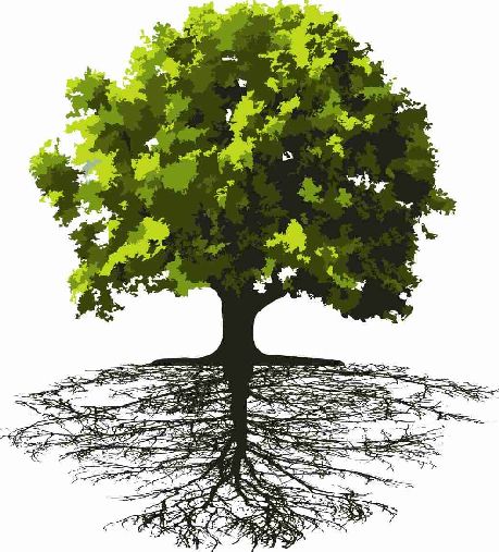
The enthesis is inserted to the bone like the roots of a tree are anchored to the ground.
This allows for firm attachment and is key to understanding the link between diseases of the enthesis and the underlying bone.
 |
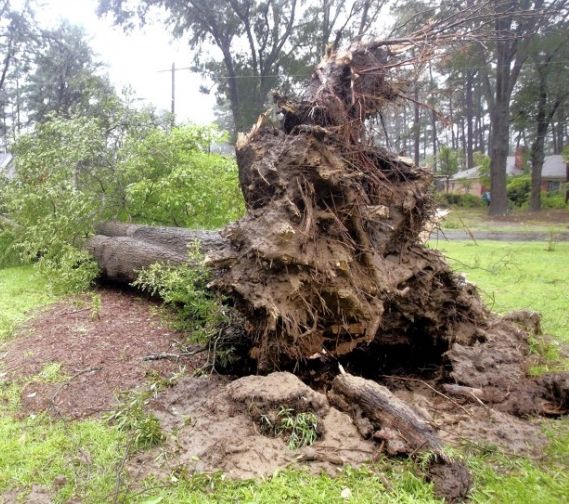 |
| The anchorage of a tree to the ground is complex with the underground root network being quite extensive (shown on left). This spreads the stress related to anchorage over a wide area. When a healthy tree is blown over in a storm the roots are uprooted but the trunk may still be intact (shown on right). | |
Sometimes the roots of a tree can be seen on the surface of the ground.
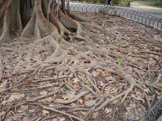 |
| This shows a tree root network that is on the surface of the ground. |
So it is with the enthesis where anchorage may be along the bone lining that is called the periosteum. This may be another cause of bone pain related to the insertions superficial anchorage to the periosteum.
Structure of the enthesis bone connection
Now turning to the analogy of an enthesis to the roots of a tree.
The figure below is a high resolution x-ray of a slice of bone taken from a knee joint at the attachment of the anterior cruciate ligament (ACL). The X-ray does not show the soft tissue of the enthesis insertion but this is drawn for explanation.
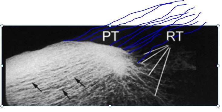 |
| From the insertion bone struts known as trabeculae can be seen running a considerable distance into the bone in the manner like a tree being anchored to the ground. PT = Patellar tendon. RT = shows the location of the anchoring bone struts that are called trabeculae. The black arrows also show the anchoring bone struts that run away for the attachment site on bone surface. |
Bone fracture related to the enthesis
So it is with human insertions where an elaborate attachment to the underlying bone plays a role in pathology. This is illustrated below for a stress fracture in the heel that is related the Achilles enthesis.
As a result of this anchorage the failure of an enthesis may manifest in the underlying bone as a fracture or microfracture.
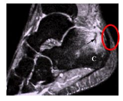 |
This is an MRI scan of an athlete who sustained a severe heel injury or stress fracture that is related to the Achilles enthesis. The MRI shows a fracture in bone adjacent to the attachment (black arrow). The actual insertion point (inside red circle) is not damaged. Black arrow = stress fracture. C = calcaneus. Red circle = Achilles tendon enthesis the anchoring bone struts that run away for the attachment site on bone surface. |
Microscopic anchorage of enthesis to bone
At the microscopic level bonding between the soft tissue of the tendon or ligament and the underlying bone is shows how this is formed.
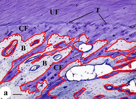 |
| This is a microscopic picture of how the insertion is connected to the bone. The tidemark (T) is the point where the shock absorbing fibrocartilage (UF) abruptly takes on the hardness of bone (CF = calcified fibrocartilage). This is then locked into the underlying bone struts (trabeculae) like a tiny network of roots (red lines added for explanation) |
The implications of this anchorage system between the enthesis and the bone is that diseases of the enthesis are associated with bone damage. This might be due to mechanical stressing of the bone and also due to bone micro-cracks and microdamage.
References
