The Enthesis Organ
Introduction
An organ is group of tissues that work together to carry out a shared function. The function of the enthesis organ is to provide a stable anchorage to the skeleton and to minimise damage at the insertion site which is subject to high levels of physical stress.
The enthesis organ
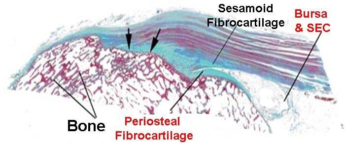
The enthesis organ is composed of the enthesis insertion and adjacent tissues. Whilst the insertion provides firm anchorage to the underlying bone the enthesis organ comprises several other tissues. The actual point of insertion is shown by the arrowheads in the figure below.
|
| This is a diagram of the Achilles tendon enthesis organ. The arrowheads show the point of attachment of the tendon to bone. The enthesis is closely integrated into the bone. Additional shock absorbing fibrocartilage termed periosteal fibrocartilage (which lines the bone) and sesamoid fibrocartilage (which lines undersurface of the tendon) are shown in sea blue. The bursa forms part of the enthesis organ. The bursa is lined by synovium which nourishes the fibrocartilages. This component of the enthesis is called the synovio-entheseal complex. |
Enthesis Organ components
The underlying bone - insertions are anchored to the bone like the roots of a tree are anchored to the ground. As a result diseases associated with the enthesis organ may be maximal within the bone.
The adjacent bone surfaces- Insertions may run over bony prominences termed tuberosities. As a result the tendon that is located well away from the attachment site proper is compressed against the adjacent bone during movement. This limits stress at the actual insertion point. A variation on this is that entheses may arise from pits of depressions in the bone leading to bone stressing being spread over a wide area.
Tendon or ligament- immediately adjacent to the attachment site- The bone and tendon surfaces adjacent to the enthesis are lined by fibrocartilage. The cartilage lining the bone surface is termed the periosteal fibrocartilage and the one lining the under surface of the tendon or ligament is called a sesamoid fibrocartilage.
soft tissues including the SEC - the cartilages related to the enthesis organ that line the bone derive nourishment from a specialised tissue called synovium. This forms a structure called a synovio-entheseal complex (SEC). This structure forms a cavity called a bursa
Adjacent supportive tissue or fascia - many insertions around the body are laden with fat which is a liquid and shock absorber at body temperature. The supportive tissue around the enthesis is called fascia and carries blood vessels that supply the enthesis. Many insertions are connected to adjacent insertions by a band fascia.
Purpose of the enthesis organ
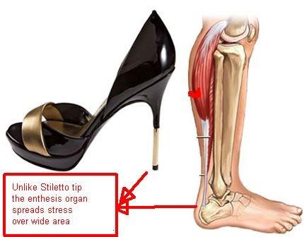
If all of the stressing was concentrated at the attachment point to bone lots of damage would occur. This is analogous to the stress a stiletto shoe heel tip exerts on a wooden floor. This is damaging to the floor in comparison to a broad based cushioned sole of a running shoe. The enthesis organ cushions and protects attachment sites to bone.
|
| The force exerted by large muscles is concentrated at the small area of insertion to the bone. The example given shows the Achilles tendon enthesis. The large forces experienced at this location can be likened to the force exerted by the tip of a stiletto shoe. To minimise the chances of damage at insertions the enthesis has several adaptations and forms a structure called an enthesis organ. |
Implications
The entheis organ is key to understanding the extensive nature of soft tissue swelling or bone abnormalities that accompanies diseases of the enthesis.
References
