The Enthesis and Bony Pulleys
Overview
Diseases of the Enthesis are often associated with an abnormal reaction in the bone at a location that is well away from the actual insertion site.
|
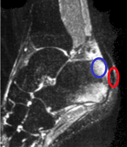
| This image shows inflammation well away from the Achilles tendon insertion. On Magnetic Resonance Imaging this pattern is termed "bone oedema". The red circle is the actual point of insertion of the Achilles tendon to the bone. The blue circle shows the "bone oedema" or inflammatory reaction within the bone. This occurs because the tendon is running over the curved bony surface or bony pulley near the attachment site. |
Why the Enthesis is associated with adjacent bone pathology
Most entheses throughout the body either run over a bony prominence called a tuberosity or are attached to a depression or "pit" on the bone surface.

|
| Example 1 shows the Achilles tendon insertion which is running over a rounded bony prominence on the calcaneus called the calcaneal tuberosity. Example 2 shows a similar arrangement in the shoulder where the supraspinatus tendon curves over the rounded surface of the humeral head or funny bone head. Example 3 is a tendon attachment in the knee called the Popliteus. In this case the enthesis is attached into a depression or pit in the bone. Example 4 is the attachment of an internal ligament of the knee called the anterior cruciate ligament. This is running over a bony prominence. Example 5 is a tendon attachment in the finger which is running over the thickened rounded end of the joint. Example 6 is a tendon on the top of the foot called the tibialis anterior that is likewise running around the curved surface of the adjacent bone. |
An Explanation for this complex anatomy
All of the arrows point to shock absorbing cartilage on the bone surface adjacent to the attachments.
The tendons or ligaments wrap around these bony pulleys and as a result the forces are spread over a wide area. This lessens stress and damage at the actual insertion points.
However pain and disease can arise in these other locations within the enthesis organ.
Implications of Bony Pulleys
In health the cartilage or fibrocartilage lining the bony pulleys prevents the tendon from 'sawing' through the bone.
In disease this anatomical structure of the enthesis contributes to the following processes:
- tendinopathy that occurs away from attachment site
- bone bruising, bone erosion and bone oedema on magnetic resonance imaging and X-ray or ultrasound
- bone surface pain termed periostitis
- this may contribute to poorly localised bone pain
You can study these processes in more detail elsewhere on this site.
References
