Bone Erosion at Normal Insertions
Introduction
The purpose of this page is to show how the normal skeleton develops microscopic erosion immediately adjacent to enthesis. This is due to microdamage and usually repairs in healthy subjects. The importance of this and the need to avoid a wrong diagnosis that such erosion is due to joint inflammation is discussed.
Mechanism of Erosion at Normal Insertions
Bone erosion is a process whereby the surface of a bone (the bone cortex) is degraded or eroded and is most typically seen in the setting of inflammation. However, the normal skeleton appears to be riddled with microscopic erosions. The enthesis is a highly mechanically stressed site which leads to microtrauma to the immediately adjacent bone. This is the basis for small erosions in the normal non-diseased skeleton which likely repair spontaneously.
Microscopic erosion at normal insertion
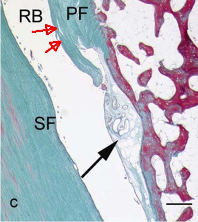
The early phases of erosion may start to damage or loss of the shock absorbing fibrocartilage that covers the bone.
|
| This is an example of microscopic erosion of the fibrocartilage of a normal Achilles
enthesis (erosion at site of black arrow). The rule is that the erosions tend to occur
at sites of predominant compression.
RB= Retrocalcaneal bursa
SF= seasamoid fibrocartilage
PF= remaining periosteal fibrocartilage or bone line tissue (red arrows)
|
Microscopic Erosion at Synovio-entheseal Complex in hand joints
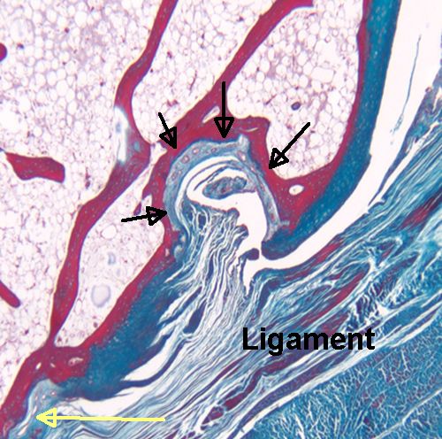
Normal small joints tend to develop microscopic erosions at sites where the ligament immediately adjacent to the enthesis compresses the bone [1]. This is because the shape of the bone leads to the forces being spread over a wide area that contributes to damage. This occurs at a structure termed a synovio-entheseal complex.
|
| The black arrows show a microscopic erosion over a knuckle joint. The overlying ligament is shown. The yellow arrow shows the point of ligament attachment closest to the joint cavity. Small blood vessels in the base of the erosion are likely linked to attempted repair. |
Small bone cysts underneath Synovio-entheseal Complex in hand joints
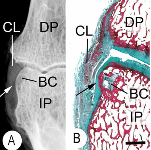
Sometimes the bone compression by the enthesis organ transmits stresses to the underlying bone and this initially manifests as a small cyst. Later on the roof of this may cave in leading to erosion.
|
| This is an X-ray (A) and a corresponding tissue section (B). It shows a small bone cyst (BC). This is underneath the cartilage lining the side of the bone. (black arrow). Damage occurs here because the ligament (CL) presses against the bone as it runs between the joints. |
Implications
These small erosions are not seen on conventional X-rays. However, they may be evident on MRI or ultrasound or microCT scanning [2].
These could be wrongly diagnosed as being related to inflammation and patients could get expensive but inappropriate therapy. This could happen when doctors scan joint with modern high technology MRI and ultrasound techniques.
References
