New Bone Formation at Normal Entheses
Introduction
The normal enthesis has a tendency to develop new bone formation with ageing. This manifests as incidental "bony spurs" that are visible on X-rays that are usually being done for another reason e.g. looking for fractures. Sometimes the new bone formation at normal entheses can be quite extensive. This page explains why this occurs and why it does not necessarily need treatment.
Basis for New Bone Formation at Normal Entheses
New bone formation occurs at the outermost part of the normal enthesis in the Achilles tendon, plantar fascia and many other sites. Joint tissues under tension favours bone development and the outermost part of the enthesis is subject to predominant tension (pulling force).
In animals when cartilage is under tension this can also lead directly to new bone. This is termed chondroid metaplasia [1]. This type of new bone may contribute to fracture repair using a surgical technique called bone distraction. This type of reaction is also well reported at the human enthesis in the healthy state [2].
Various molecules known to be key to new bone formation include bone morphogenetic protein-2 (BMP-2) and BMP-7 are up regulated by mechanical stress [3].
|
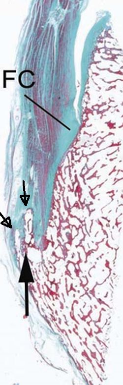
| This is a normal aged Achilles tendon enthesis. The black arrows show new bone formation on the outside of the enthesis. Where bone is under tension or being pulled creates an environment that favours new bone formation. This occurs normally with age but is accelerated in disease states such as Ankylosing Spondylitis and Psoriatic arthritis. FC = fibrocartilage |
Implications and Pitfalls
The new bone formation that occurs in diseases such as Ankylosing Spondylitis and Psoriatic Arthritis is an exaggeration of the normal tendency to make bone on the outer surface of the enthesis.
Switching off inflammation in Ankylosing Spondylitis does not change the mechanical environment of the enthesis. Therefore, new bone formation may continue at the previously inflamed level.
A bone spur in the name applied to enthesis new bone formation. It is termed a spur as it is thicker at the base and tapers. Pain at the enthesis is often attributed to bony spurs when these are present or visible. Removal of spurs in the majority of cases may not make much difference.
New bone formation may occur from a previous injury related to bleeding near the enthesis. It is important to understand that new bone at the enthesis may not be the cause of pain and its removal may not necessarily be needed. This is a last resort to try and alleviate pain.
References
