The Enthesis Insertion
Overview
The images in this section show the actual insertion of a fibrocartilagenous enthesis to the bone as viewed microscopically using a stain called Masson's Trichrome.
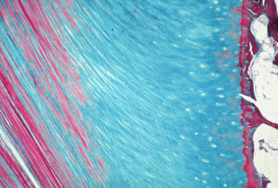 |
| The red vertical lines on the left are collagen tendon fibres. The blue colour in the middle of the image is the fibrocartilage [link]. The red area is the transition zone from fibrocartilage at the attachment site. The black box shows the actual area of attachment which is magnified in Figure 2 below. The white on the far right of image is bone cavity fat. |
 |
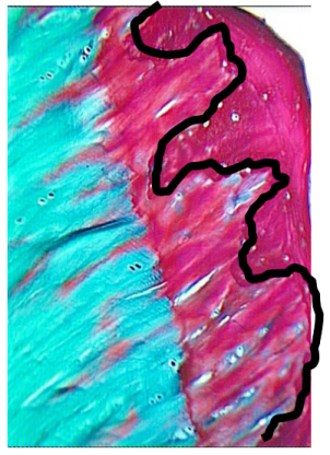 |
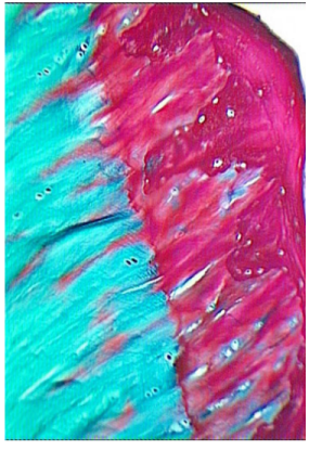
| Figure 2s and 2b are identical. Fig 2a shows an abrupt transition of fibrocartilage in blue to calcified fibrocartilage in red. At this "tidemark" there is an abrupt transition from soft compressible cartilaginous tissue to bone hard tissue. | A black line in Figure 2B shows the "jigsaw like" interlocking between the calcified fibrocartilage and the underlying bone. This thin line anchors the soft tissue to the underlying skeleton. |
Therefore this region is composed of:
- Tendon or ligament
- Fibrocartilage
- A tidemark
- Calcified Fibrocartilage
- Jigsaw like locking into the bone
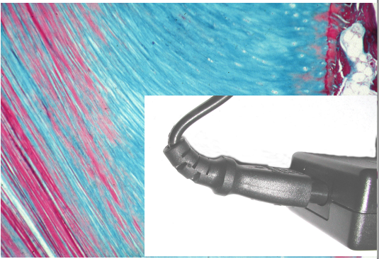 |
| The fibrocartilage acts like a grommet on an electrical cable by protecting the cable from damage at the insertion to the plug. The actual insertion is beneath this region and is interlocked like a jigsaw but is protected by the overlying tissue. |
Things go wrong
The images depicted above show things in a state of health. However, the high stressing and damage and disordered repair at the enthesis-bone interface is a major cause of arthritis with pain and loss of function. This is covered elsewhere on this website.
References
J Anat. 2001 Nov;199(Pt 5):503-26. The anatomical basis for disease localisation in seronegative
spondyloarthropathy at entheses and related sites. Benjamin M, McGonagle D. 