The Plantar Fascia Enthesis
Introduction
The plantar fascia is a fairly well known enthesis that is often associated with the painful heel condition known as plantar fasciitis. It is the largest enthesis in the body in an orientation that runs parallel to the ground. Most other large entheses are perpendicular to the ground. This configuration could be very important for understanding pain related to degenerative conditions of the plantar fascia. This page describes the anatomy of the plantar fascia.
Basic Anatomy
The plantar fascia is a thick band of tissue that is inserted to the under surface of the heel bone (calcaneus). It runs forward to the base of the toes and forms the medial arch on the foot. This contributes to optimal foot mobility and the "spring" of the foot.
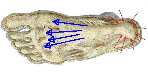
|
| This is an anatomical dissection of the plantar fascia. The fairly extensive distribution of the attachment site is shown by the red arrows. The fascia runs forward to the base of the toes and is shown by the blue arrows. Tension along this structure helps form the arch of the foot. |
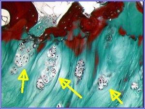
Tissue Structure
The plantar fascia is a typical fibrocartilagenous enthesis. The undersurface of the plantar fascia is associated with a considerable degree of bone compression. This is associated with new bone formation (termed spurs) that is a normal age related change.
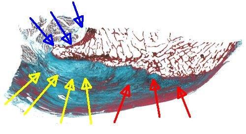
|
| This image shows the microscopic structure of the normal plantar fascia. The red arrows show the fibrous part of the plantar fascia. The yellow arrows show the fibrocartilage which is most abundant at sites of compression. The blue arrow shows the development of new bone (spurs) adjacent to the deepest part of the plantar fascia. |
New bone at the plantar fascia termed bony spurs is commonly seem on X-rays.
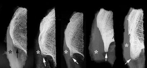
|
| These 5 images show bony spurs of different sizes at the plantar fascia. The spurs are progressively larger from left to right. The asterisks show the actual enthesis. The arrows show that the spurs are deep to the enthesis and are not actually part of the insertion. Rather these spurs help distribute the load from the plantar fascia when it is being compressed against the ground. |
These spurs are not at the enthesis but are adjacent to it. The distribution of the spur formation is similar to the distribution of new bone formation in osteoarthritis (OA) which is termed osteophytes. In OA these typically occur at the joint margin rather than at the enthesis.
Most spurs at entheses arise at sites of tension. The compression of the plantar fascia against the ground is associated with bone formation at the margin of the enthesis.
Microdamage
As for entheses elsewhere, the high stresses at the plantar fascia is associated with tissue microdamage. This likely undergoes repair throughout life and likely occurs to a variable degree in everyone. Why some people get pain at this site and others do not is unclear, but may link to the actual degree of microdamage.
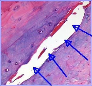
|
| This image shows a microscopic crack in the fibrocartilage of the plantar fascia (blue arrows). This is a feature that is typical of wear and tear at other sites in the skeleton. |
|
| This image shows that the chondrocytes (cartilage producing cells) at the enthesis have formed proliferating nests that are typically seen in the repair reaction that accompanies "wear and tear" or osteoarthritis at other sites in the skeleton. |
References
