Enthesis Blood Supply
Introduction
The actual enthesis point of insertion to bone lacks a blood supply and is described as avascular. The specialised shock absorbing tissue called fibrocartilage is found at this location. This page explains the nature of the blood supply in the vicinity of the enthesis.
The Enthesis Blood Supply
The fibrocartilage at the enthesis has a very low oxygen requirement. Air contains 21% oxygen. The oxygen tension in fibrocartilage is thought to be from 1% upwards. Oxygen gets into the various parts of the insertion region by diffusion. The tissues adjacent to the insertion that form part of the enthesis including ligaments or tendons also have a comparatively low density of blood vessels.
The fibrocartilage "steals oxygen" from the adjacent tissues including the bone and tendons or ligaments and local supporting tissues called fascia. Synovium that is part of a specialised structures called the synovio-entheseal complexes also nourishes fibrocartilage via synovial fluid.
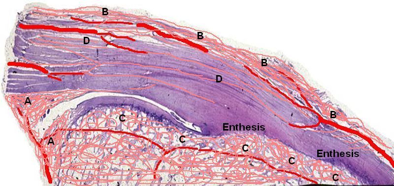
The blood supply of an enthesis is drawn in red in cartoon form below.
Studies in living subjects confirm that the attachment site lack a blood supply. Blood vessel approach the enthesis from the bone marrow, the substance of ligaments or tendons and the tissue outside the enthesis.
|
| A = Synovio-Entheseal Complex which is well supplied with blood vessels. B = The peritenon or surface of the tendon. Ligaments have the same configuration. C = The blood vessels in the bone beneath the enthesis. D = Shows the very low density of vessels in the tendon near the enthesis. It also shows that the actual attachment site to bone that is termed fibrocartilage has no vessels at all. |
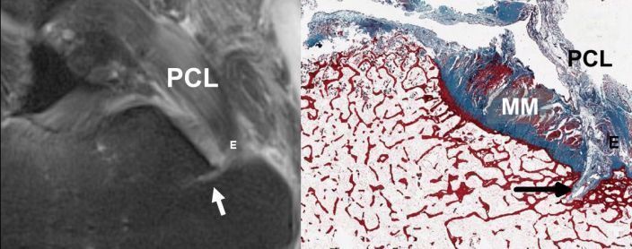
An Important blood vessels closely related to the Insertion.
Many entheses have a blood vessel that in situated underneath the part of attachment site that moves the least during joint motion.
Where this blood vessel enters the bone creates a potential tunnel for inflammation within the synovio-entheseal complex to spread to the bone.
|
| The picture on the left is a knee MRI scan. PCL = Posterior cruciate ligament. E = enthesis. The white arrow shows the blood vessel at the base of the enthesis running into the bone. The image on the right shows the corresponding location of the blood vessel in the actual tissue from the knee. |
Implications
Tissue microdamage at the enthesis leads to a need for tissue repair to stop the enthesis failing.
The downside of the low degree of blood supply to the enthesis might be very slow or inadequate healing responses, especially with age. This may be a major contributory factor to chronic pain and disease.
The upside of the lack of blood vessels at the enthesis is that this site is partially protected from inflammatory reactions or "immuneprivileged" The very low density of blood vessels makes it very difficult for immune cells to get near the attachment site.
With injury to the enthesis blood vessels can penetrate the avascular sites to carry out the tissue repair. This might be a key event in immune activation in chronic enthesis disorders such as Ankylosing Spondyliits
In inflammatory diseases the fibrocartilage may be destroyed and blood vessels invade this region. What happens to the blood vessels in diseases of the enthesis will be addressed in the section dealing with disease.
References
