Does Nail Related enthesopathy play a role in pincer nails?
Nail Anatomy
The outside of the nail was thought to be anchored to the skin.
Surgical dissection work and recent tissue histology studies have shown that the outside of the nail (lateral nail) is actually anchored to the ligament attachment sites.
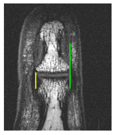 |
This is a Magnetic Resonance Imaging scan of the distal interphalangeal joint and the nail region. The yellow line on the left indicates where doctors originally thought that the strong anchorage of the joint called the collateral ligament actually was placed. However, when we looked in detail it was evident that the ligament ran along the nail to help tether its edges down. |
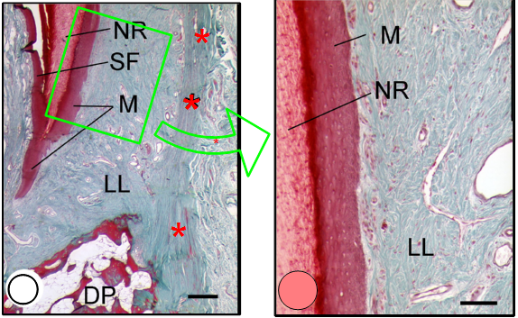 |
| This is a microscopic view of the outside of the nail. The red asterisks show the course of the ligament that runs up along the nail. The region called the lateral lamina is thick connective tissue that tightly anchors the nail to the ligament. |
How disorders of lateral tethering might cause nail problems
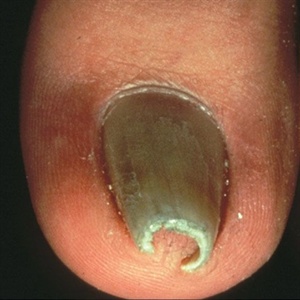
Excessive curvature of the nail plate may lead to the nail developing a dome like curvature.
This gives an appearance of the nail being pinched, or so called pincer nails.
A pincer nail, also called a trumpet nail, is a type of an ingrown nail which can be quite painful.
It can occur after entities such as fungal infections. It most typically afflicts the big toes.
It may require surgical treatment including complete removal of the nail to prevent recurrence.
The cause of pincer nails is poorly understood. We believe that a dysfunction in the integration of the nail with the collateral ligament enthesis may play a significant role in this condition.