Costochondritis as a Cause of Undiagnosed Chest Pain
Overview
The attachments between the ribs and the breastbone are called costochondral joints.
These insertion points may be associated with the development of chest pain.
This page provides a case history of such a patient and also an anatomical explanation.
An Illustrative Case
A 40 year old man was admitted to the Coronary Care Unit of the Hospital with severe central chest pain.
The pain came on after exertion and was associated with breathing difficulties or shortness of breath.
The patient had a strong family history of heart disease with his father passing away aged 45 with a myocardial infarction or heart attack.
The patient was also a smoker.
He was slightly overweight and admitted to being under severe stress at work.
The admitting doctors suspected that he might have coronary artery disease.
An extensive array of tests were performed including blood investigations, ECG, Cardiac Ultrasound and a coronary artery angiogram.
All of these were normal.
A diagnosis of angina pectoris was made and the patient was treated with low dose aspirin and a nitrate patch and discharged from hospital.
In the following 3 months he was admitted 4 times to hospital with suspected acute coronary insufficiency (partially blocked coronary arteries).
Over that time he spent 2 weeks in the Coronary Care Unit.
He also had investigations for upper gastrointestinal disease and also had a trial of anti-acid reflux therapy without benefit.
Likewise there was no evidence for pleurisy or other lung problems.
The treating Consultant physician noted that the patient had some tenderness adjacent to the breastbone.
Rheumatology opinion was sought.
The Rheumatologist noted that the pains were fairly constant throughout the day.
The Rheumatologist noted that there was no personal or family history of Psoriasis or Colitis or inflammatory arthritis. Therefore the pain was more likely "Mechanical" rather than "inflammatory".The patient had no pains elsewhere.
The patient was tender along the left edge of the breastbone with some focal tender spots.
There was no swelling evident.
A diagnosis of costochondritis was made.
The two most tender costal cartilages were injected with corticosteroids.
An accurate diagnosis had a very positive impact on the patient who now felt less distressed since the prognosis in costochondritis is much better than heart disease.
The patient was discharged the following day and was seen 3 months later in the Rheumatology Outpatients where he still got some discomfort in the chest but was much better.
The anatomical basis for Costochondritis
 |
|
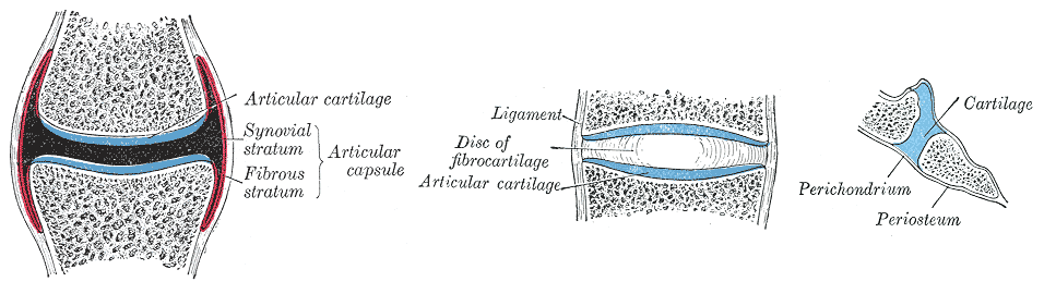
This cartoon shows a synovial joint on the left. These are typically found in the knee and hand
joints for example and permit a good range of movement. The articular cartilage is in blue. The middle
figure shows a vertebral disc joint. Cartilage and fibrocartilage are in blue. This type of joint
allows a degree of movement. The image on the right is a cartoon of a costochondral joint. The blue
represents a cartilaginous bridge between the two ends of the bone. Such joints have very little movement.
|
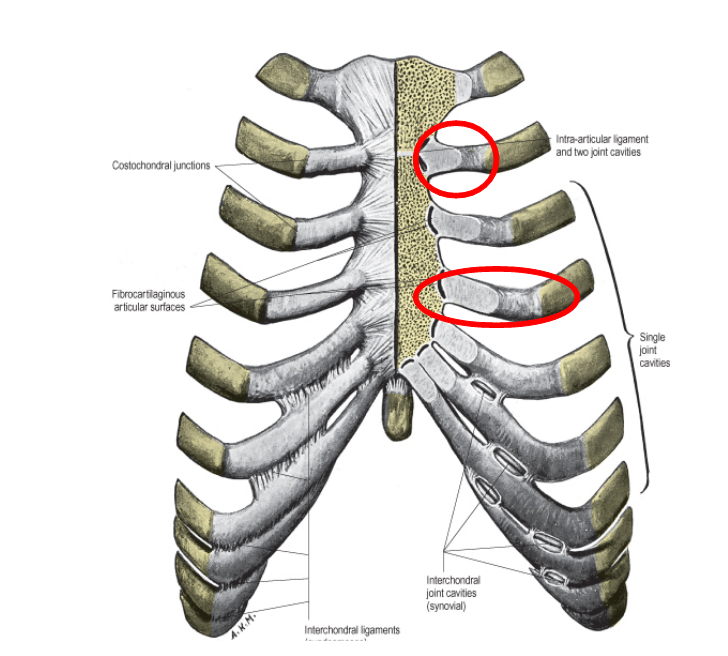
As shown in the figure below some of these joints have a small synovial cavity that allows limited movement in one plane.
 |
|
This diagram shows the costochondral joints in the breast bone. Rib joint 2 to 7 have small synovial
cavities in the joints. The right hand side of breastbone is cut down to show the joints. The lower
costochondral cartilages are joined to each other. With age the costochondral joints can covert completely
from cartilage to bone. The second and fourth costochondral joints are displayed inside the red circles
|
Learning Points
Chest pain is a potentially serious condition so patients should always seek medical advice as soon as possible.
If you suffer from undiagnosed chest pain then costochondritis is a possibility but other more serious conditions first may need excluding.
Costochondrits is thought to be a type of inflammation where the ribs join the breastbone or what is termed an enthesopathy. There is a lack of tissue studies (termed histology) of costochondritis.
The examination in a patient with costochondritis may be normal. A tear adjacent to the insertion of one of the rib muscles or a "pulled muscle" is also a possibility.
You can learn about disorders of the enthesis or insertion points on this website.