Fat at the Enthesis
Introduction
Fat or adipose tissue is intimately associated with normal enthesis function. Fat is a shock absorbing material at or adjacent to the enthesis. The fat tissue at the enthesis has lots of nerve endings and may play a very important role in pain.
Relevant Anatomy and physiology
At body temperature fat is a liquid. This allows for easy movement and permits fat to act as a space filler at the enthesis. It is held within cells which in turn are held within structures called fibrous septae that anchor the fat in the vicinity of the enthesis. This allows fat to cushion the enthesis.
Locations of Fat at the Enthesis
The locations where fat is found are listed below
Fat pads. The heel has a large fat pad that protects the plantar fascia enthesis. A small fat pad or an abnormal fat pad at this site is associated with a greater propensity for plantar fasciitis or diffuse vague heel pain.
|
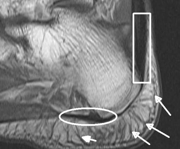
| This is a MRI scan of the heel. Inside the white box is the Achilles tendon insertion. The round white oval shows the attachment site of the plantar fascia. The white arrows show the bands of tissue called fibrous septae that hold the fat pad in place. In health this prevents it being dislocated during movement. This configuration is common throughout the body and helps protect the enthesis. |
Fat within bursae. Many enthesis form structures termed synovio-enthseal complexes. These structures are lined by synovium which nourishes the enthesis related fibrocartilage. Fat pads are located under the synovial lining.
|
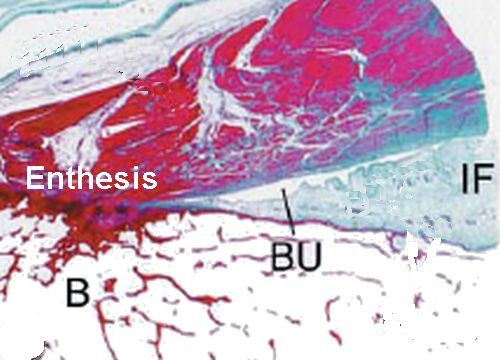
| The region forms a space called a bursa (BU). A large fat pad (IF) is evident within the bursa. Such fat pads move in and out of the bursa during movement. This acts as a plunger and stops tissues knocking against each other. B= bone. |
Fat within tendons. Several large tendons have fat within their substance as they splay out to attach to the bone.
|
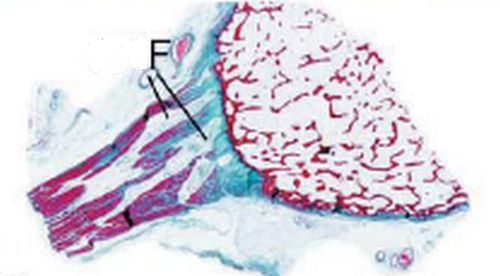
| This image shows fat within an insertion region where it splays out to insert into the bone. The tendon fibres splaying leads to the space void being filled by fat |
Fat on surface of tendons. The peritenon, or surface of the tendon may have a layer of covering fat.
|
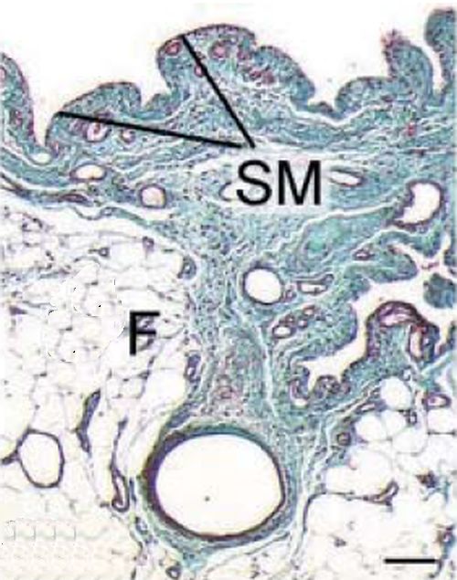
| The surface of many entheses is covered with fat immediately adjacent to the attachment site to bone. This image shows the synovium (SM) lining the fat (F) that covers the surface of the anterior cruciate ligament of the knee. This facilitates smooth movement of the ligament and enthesis |
Other functions of fat at the enthesis
The fat at the enthesis has a rich nerve supply. This helps in sensing joint position and may also be important for pain.
The fat at the enthesis is rich in immune cells termed macrophages. This could be very important in contributing to inflammation at the enthesis including inflammation at synovio-entheseal complexes.
Implications
Damage or injury to fat pads at the enthesis may be associated with plantar fasciitis. Damage to the fat pad deep to the patellar tendon enthesis may be associated with pain in the front of the knee. The affected knee fat pad is known as Hoffa's fat pad.
References
