Fibrocartilage Microdamage
Introduction
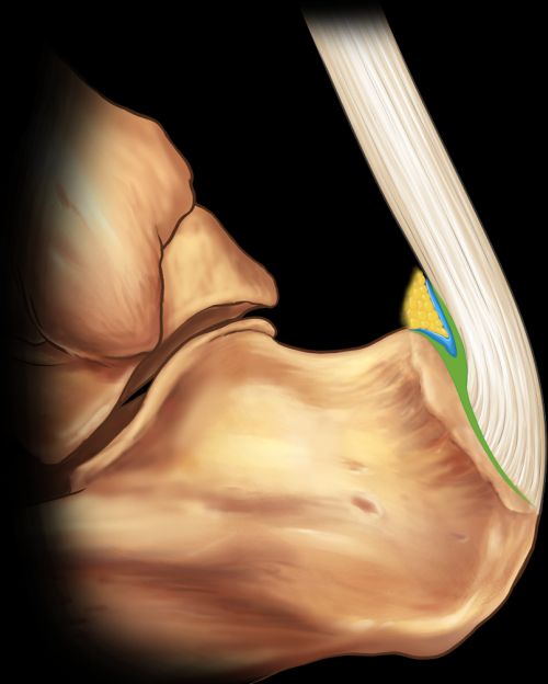
Fibrocartilage is the shocking absorbing tissue at the enthesis insertion site and within the adjacent synovio-entheseal complexes. The high forces being exerted at the fibrocartilage makes it prone to microscopic damage termed microdamage. The extent of the fibrocartilage distribution at the Achilles tendon enthesis organ is shown in the figure below.
|
| The green area shows the extent of shock absorbing fibrocartilage. The blue region shows the synovio-entheseal complex tissue that is prone to becoming inflamed. Damage to the fibrocartilage may trigger inflammation and pain in this region. Image courtesy of Prof Sibel Aydin |
Fibrocartilage Microdamage
Fibrocartilage microdamage is very common with normal ageing [1]. At what age this fibrocartilage damage starts is not clear. It is likely that microdamage in younger subjects quickly repairs but in older subjects this repair process is less efficient.
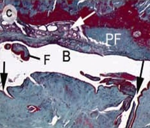
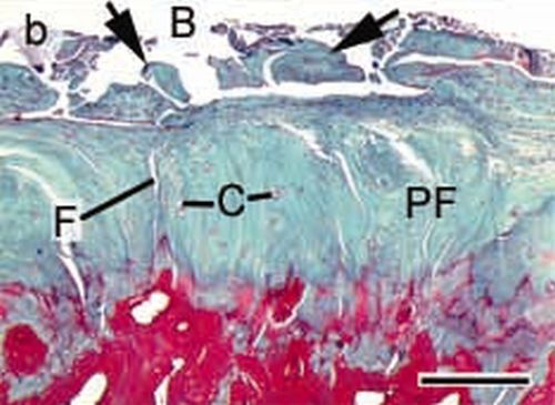
The images below illustrate enthesis fibrocartilages damage which is very common in the normal aged skeleton. The fibrocartilage should be smooth and regular but is irregular and cracked due to microdamage
|
| This image shows extensive fibrocartilage microdamage. The black arrows show deep clefts in the damaged fibrocartilage. F = fragment of fibrocartilage that has broken off |
|
| The arrowheads show fragmentation and breakage of the fibrocartilage lining F= fissures of deep cracks in fibrocartilage PF= periosteal fibrocartilage which means the type that lines the bone. Bone is red underneath |
Implications
It is well known that fragments of damage tissue can elicit powerful immune response that may contribute to inflammation and pain.
Excessive fibrocartilage microdamage in athletes may contribute to enthesis inflammation and pain. This is poorly understood at present.
The decline in fibrocartilage repair capacity in older subjects is likely to be a major factor contributing to joint degeneration in osteoarthritis [2]
References

Resources
There is very little information on fibrocartilage damage.
University of Leeds Histology Guide Cartilage: The three types of cartilage