Nail Anatomy and The Nail Enthesis
Overview
The nail is a tough hard shield on the extremities of the fingers and toes.
It has several functions including:
- protection of the digits from trauma
- a role in fine movements and pinching
- a role in sensory perception
- an emerging understanding of its role in skeletal function
The structure of the actual nail is well reported but the amazing fact that the nail is anchored to the skeleton has only recently been fully appreciated.
This anchorage is crucial for understanding nail pathology and for surgical treatments for various nail disorders.
This page focuses on the nail root or nail matrix region which is the site the nail originates.
|
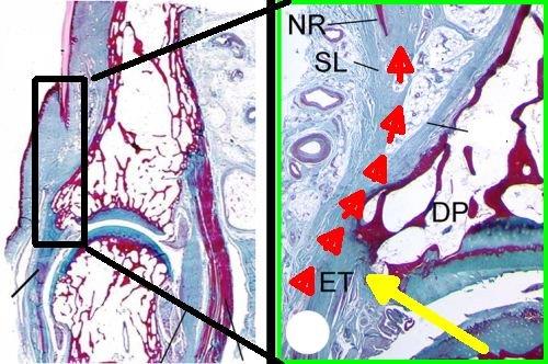
| The left panel shows the nail and the distal interphalangeal joint (DIP) on low power microscopy. The image on the right is a magnified one of the actual DIPl tendon attachment to the bone and its link to the nail. The yellow arrow shows the DIP tendon enthesis. The series of red arrows show how the tendon does not completely attach to the bone but anchors directly to the nail as shown by the red arrowheads (NR= nail root). This means that the nail is directly connected to the skeleton. |
In 1956 Dr Verna Wright noted a strong but inexplicable link between nail disease and Psoriatic Arthritis [1]. [see also Enthesitis, Enthesopathy and the Nail in Psoriatic Arthritis]
The discovery by our team that the nail was anchored to the skeleton was a real cliffhanger [2]. It provided and explanation for both arthritis and mountaineering!

References
