Enthesitis Causing Knee Joint Swelling in PsA
Knee Disease in PsA can occur as part of a generalised form of PsA or may be confined to the knee. The knee is a large joint with over 30 entheses within or immediately adjacent to the joint.
Viewing Enthesitis in PsA related Knee Disease
Historically, X-ray studies have shown occasional new bone formation at the entheses in knee joint disease in PsA compared to RA. The initial use of MRI in PsA showed no difference between it and rheumatoid arthritis (RA). Work in Leeds reported in 1998, showed that enthesitis and osteitis were commoner in PsA related knee disease than RA [1].
|
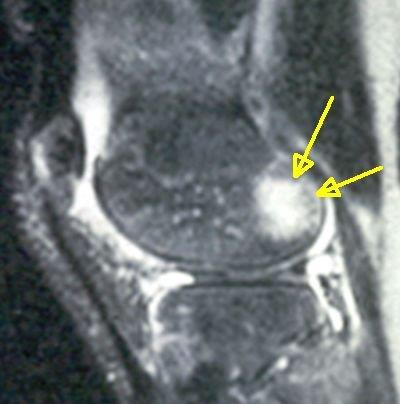
| in patients with early knee rheumatoid arthritis (RA) showing joint fluid accumulation (effusions) due to joint lining inflammation (termed synovitis). |
|
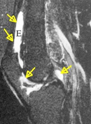
| A proportion of PsA cases have diffuse bone marrow inflammation (osteitis) adjacent to entheses (yellow arrows). Joint fluid accumulation (effusions) and joint lining inflammation (synovitis) is also evident. |
Several other studies have shown the clinically silent enthesopathy is common in the knees of psoriasis subjects. This suggests that entheseal changes may be primary lesion or the site where disease starts. This can't be proven in humans but the primary role of the enthesis has been shown in animal models.
Knee osteitis on MRI
Studies have shown that diffuse bone inflammation in the sacroiliac joint is linked to progressive joint destruction and joint fusion at that site.
However, the MRI changes in the knee were not associated with more severe or more chronic disease. The presence of skin psoriasis was the best predictor of chronicity in people with swollen knees [3]
Implications
Rheumatologist can usually recognise PsA based on the patients story, examination and blood tests. A role for routine MRI scanning has not been proven for the diagnosis of knee disease.
However, patients with knee swelling without a diagnosis often have MRI scans to look for structural damage to ligaments, menisci or cartilage. Patients having a knee MRI should have fat suppression scanning which is now pretty standard in all health care systems. Diffuse bone marrow oedema on MRI adjacent to insertions raises the possibility of PsA or a related SpA as the diagnosis.
|
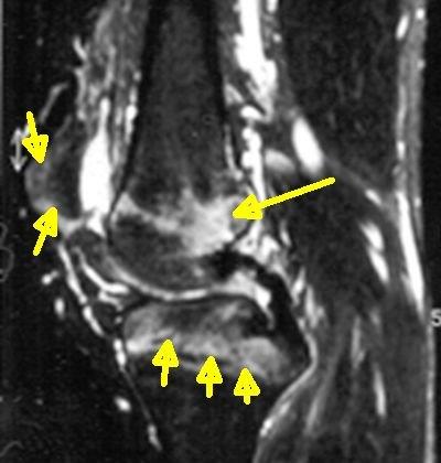
| This is an MRI scan from a 30 year old male with knee pain and swelling thought to be due to damage to the internal structure of the joint. All of the joint structures including ligaments and menisci were normal. Knee joint inflammation was evident and was linked to enthesitis on the outside of the knee joint. The yellow arrows show inflammation at knee entheses attachment sites. |
References

Resources
Knee-pain-explained.com and things to consider that leads to knee swelling Swollen knee