The Enthesis and Bone Erosion in Rheumatoid Arthritis
History of Erosion in Rheumatoid Arthritis
Bone erosion refers to the acid dissolution of bone that occurs in different types of arthritis.
Bone erosions are indicative of a more aggressive form of Rheumatoid Arthritis that needs prompt treatment.
Historically erosion in Rheumatoid Arthritis was largely considered to be driven by the immune system.
Later is was considered that joint cells called fibroblasts were key to erosion.
With knowledge of anatomy of the Synovio-Entheseal Complex it emerged that the enthesis played a pivotal role in bone erosion. The mechanism of how the enthesis drives erosion in Rheumatoid Arthritis is set out below.
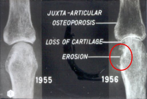 |
| Small joint bone erosion of the hands and feet is common in Rheumatoid Arthritis and is associated with a worse course of disease. This image shows the development of an erosion over a one year peroid (erosion inside red circle). |
 |
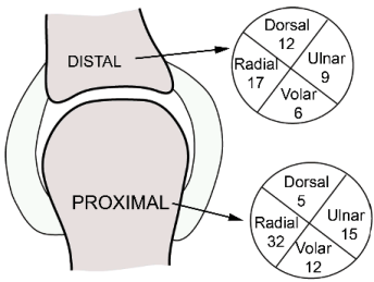 |
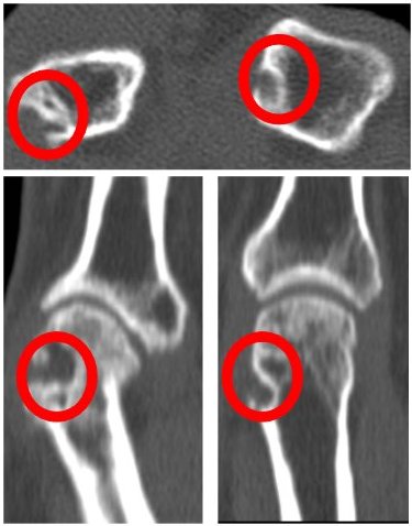
| On 3D scanning of small joints it was noted that the majority of erosions occurred near the ligament entheses. Erosions are especially common on the side of knuckle on the radial side of hand (side on which thumb is on). |
 |
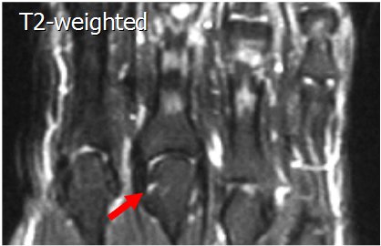 |
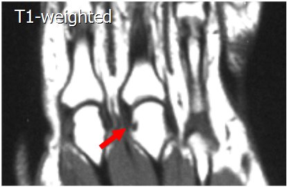
| On 3D scanning, in this case Magnetic Resonance Imaging, it is evident that small erosions can be seen in normal subjects who don't have arthritis. From Tan AL, et al. Arthritis Rheum 2003;48(5):1214-22 | |
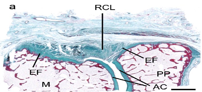 |
| This is histology of the normal small joints of the hands. The region labelled AC is shock absorbing cartilage on the end of bones. The region labelled RCL is the joint ligament. The region inside the red circle in is the ligament enthesis and adjacent Synovio-Entheseal cartilage covering the adjacent bone surface to protect it from the effect of ligament compression during joint movement. It is at this site that small erosions occur in normal subjects. McGonagle et al Arthritis Rheum 2009 |
 |
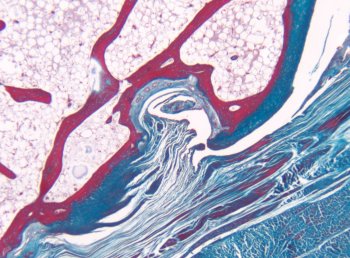 |
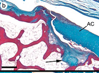
| This is a tissue specimen from a normal joint showing microscopic erosion and damage at the sites of ligament compression. McGonagle et al Arthritis Rheum 2008 | |
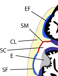 |
Summary of erosion mechanism. The collateral ligament (CL) presses on bone. Bone erosion develops at the sites of compression. The enthesis associated cartilages attempt to resist the damage associated with compression. |
Implications
Erosion formation is secondary to joint inflammation in Rheumatoid Arthritis.
It is likely that small microscopic erosions that occur in normal subjects at the enthesis grow much larger because of the joint inflammation.
Normal subjects are prone to tiny erosions at the enthesis. These heal in healthy subjects.
Control of inflammation in Rheumatoid Arthritis stops such erosions becoming larger and thus contributing to joint damage.
References

All of the figures on this page are courtesy of the images contained in these two Arthritis & Rheumatism papers.