Telling Mechanical-related, Degenerative and Inflammatory Enthesopathy Apart
Definitions
- Mechanically related enthesopathy
- typically may occur in the heel someone who walks a lot and wears hard soled shoes. An example would be "Postmans heel"
- Degenerative enthesopathy
- refers to "wear and tear" or Osteoarthritis associated disease in older subjects. This certainly overlaps with mechanically induced disease.
- Inflammatory enthesopathy or enthesitis
- refers to scenarios where the primary problem is inflammation in the insertions
Inflammatory and mechanically-related enthesopathy overview
Mechanically-related tendinopathy or enthesopathy may occur from injury, including sports-related activity.
Imaging studies may confirm the presence of entheseal pathology but the appearances, whether at the annulus or bone-disc interfaces in the spine or the plantar fascia or elsewhere, may be similar in both mechanical and inflammatory disease.
In the case of the Achilles tendon, degenerative tendon disease typically occurs 2-6 cm proximal to the enthesis itself, whereas inflammatory disease is based around the insertion and adjacent bone but there may be overlap.
Why it can be hard to always identify the cause of enthesopathy
It may be that both inflammatory and degenerative enthesopathy share common features.
For example, entheses are sites of high mechanical stressing and with age normal entheses are subject to wear and tear; thus degenerative changes similar to those seen in osteoarthritic articular cartilage may develop.
Degenerative problems may be associated with secondary inflammation.
Primary inflammatory enthesopathies (known as enthesitis) may be associated with secondary degenerative changes in some cases.
How inflammatory and degenerative disease may look similar
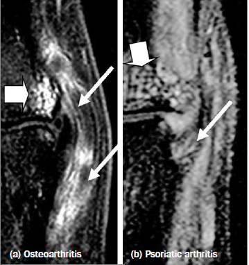
Imaging of small joints in Osteoarthritis and Psoriatic Arthritis shows that enthesis-related abnormalities are common to both conditions.
The changes are qualitatively similar in the two but quantitatively greater in the primary inflammatory disorders (Figure 1).
In fact, the similarities between mechanical and inflammatory enthesitis have highlighted the possibility that site-specific biomechanical factors may lead to disease initiation in Spondyloarthropathy.
Such factors include microdamage and repair.
It is little wonder that the differentiation between mechanical and inflammatory disease can sometimes be difficult.
In patients with chronic symptoms doctors should always consider a re-evaluation of the diagnosis.
 |
| Fig 1. The thin arrows show increased fluid in the soft tissues of the enthesis in both Osteoarthritis and Psoriatic Arthritis ("degenerative" and inflammatory respectively). The arrowheads shows a similar pattern of bone abnormalities. |