Functional Enthesis - the Tendon
Introduction
The spondyloarthropathy group of diseases localise to tendon or ligament insertion sites.
In Spondyloarthropathy, inflammation where tendons wrap around bones is also common. Tendons are lined by synovium which produces synovial fluid and lubricates the tendon. Historically it was thought that the disease process in Spondyloarthropathy started in the synovium and was not related to the enthesis.
This page explains why tendons behave like "a functional enthesis".
Different types of Tendon Pulleys
Tendons often don't just run from one location to another in a straight line but run over bony surfaces. This spreads the stress over a wide area.
The tendon may interact with the bone surface at:
- bony prominences e.g. calcaneal tuberosity of heel at the Achilles tendon
- bony grooves e.g. poplitues tendon groove
- synovial joints that often bulbous or rounded which spreads stressing forces from the enthesis to the tendon also.
- tendons must be held on the bone surface to prevent them from "bowstringing". This is done by thick reinforced bands of tissue called fibrous retinaculae that are reinforced tunnels where tendon runs through
- discrete fibrous pulleys e.g. the A1 pulley
Functional Enthesis Similarities with tendon or ligament enthesis
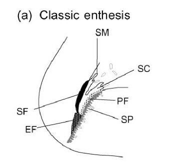
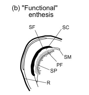
The enthesis and the functional enthesis share the following:
- similar Biomechanics
- presence of fibrocartilage
- similar tendency towards microdamage
- similar bone related abnormalities on MRI.
This is depicted in the cartoon below.
|
|
| EF = enthesis fibrocartilage (unique to insertion point). SC= synovial cavity. P = periosteal fibrocartilage. SF = sesamoid fibrocartilage. SC = subchondral plate or underlying bone. R = retinaculum which is anchoring band like tissue to stop tendon slipping off bone surface. | |
Fibrocartilage at Functional Enthesis
Fibrocartilage is present on the under-surface on tendons in contact with bone.
Fibrocartilage is also present on the adjacent bone surface and by absorbing stress prevents the tendon "sawing" into the bone and prevents the tendon wearing out on the bone.
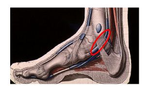
The image below shows a good example of the anatomical location of an important functional enthesis at the tibialis posterior tendon where it wraps around the ankle joint (located inside red circle).
|
EF = enthesis fibrocartilage (unique to insertion point). SC= synovial cavity. P = periosteal fibrocartilage. SF = sesamoid fibrocartilage. SC = subchondral plate or underlying bone. R = retinaculum which is anchoring band like tissue to stop tendon slipping off bone surface. |
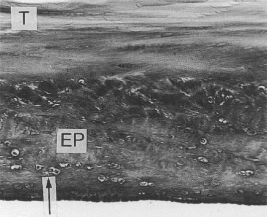
The figure below show the fibrocartilages of this same tendon functional enthesis.
|
| Image shows fibrocartilage formation on the under-surface of the tibialis posterior tendon where it wraps around the ankle. This is an adaptation to complex mechanical stressing on the surface of the tendon where it is compressed against the bone EP = epitenon or tendon surface. Arrow shows fibrochondrocyte. |
|
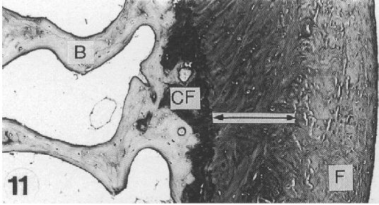
| Image shows fibrocartilage formation on the bone surface of the ankle where the tibialias posterior tendon wraps around the ankle. This is also the mechanical adaptation to complex mechanical stressing on the surface of the bone that is compressed against the tendon. |
Implications
The functional enthesis concept is key to understanding unexplained pain or swelling in the foot or ankle or wrist region.
It is important for understanding why the enthesis is linked to sausage digits or dactylitis.
The role of the functional enthesis in tendonitis and overuse injuries is overlooked
References


Further Information
http://www.nhs.uk/Conditions/Tendonitis/Pages/Introduction.aspx